
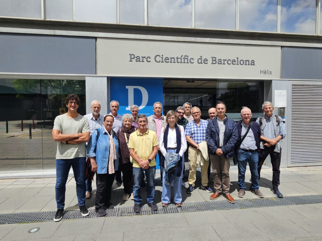
On 12 May, the Institute for Bioengineering of Catalonia (IBEC) welcomed 15 students from the Popular University of Granollers (UPG), near Barcelona, for an educational visit focused on advanced microscopy and biomedical research. The activity featured presentations and laboratory demonstrations led by Gabriel Gomila, SPM4.0 Coordinator, and Jhon Pazos, SPM4.0 Doctoral Candidate, providing participants with a unique opportunity to discover how Atomic Force Microscopy (AFM) is used to investigate biological systems at the nanoscale.
As part of its commitment to promoting scientific outreach and public engagement, the SPM4.0 project regularly participates in activities that bring cutting-edge research closer to diverse audiences. Through visits, workshops and educational events, researchers have the opportunity to share their work with students and members of the public, highlighting the role of advanced microscopy techniques in addressing biomedical challenges and fostering a broader understanding of scientific research.
With this vision in mind, SPM4.0 researchers at IBEC Gabriel Gomila (project coordinator) and Jhon Pazos, Doctoral Candidate, welcomed a group of 15 students from the Popular University of Granollers (UPG). Founded with the aim of promoting lifelong learning and public engagement with knowledge, the UPG offers a wide range of educational and cultural activities for adult learners. By connecting participants with experts from different fields, the institution fosters scientific literacy, critical thinking and a deeper understanding of the advances shaping today’s society.
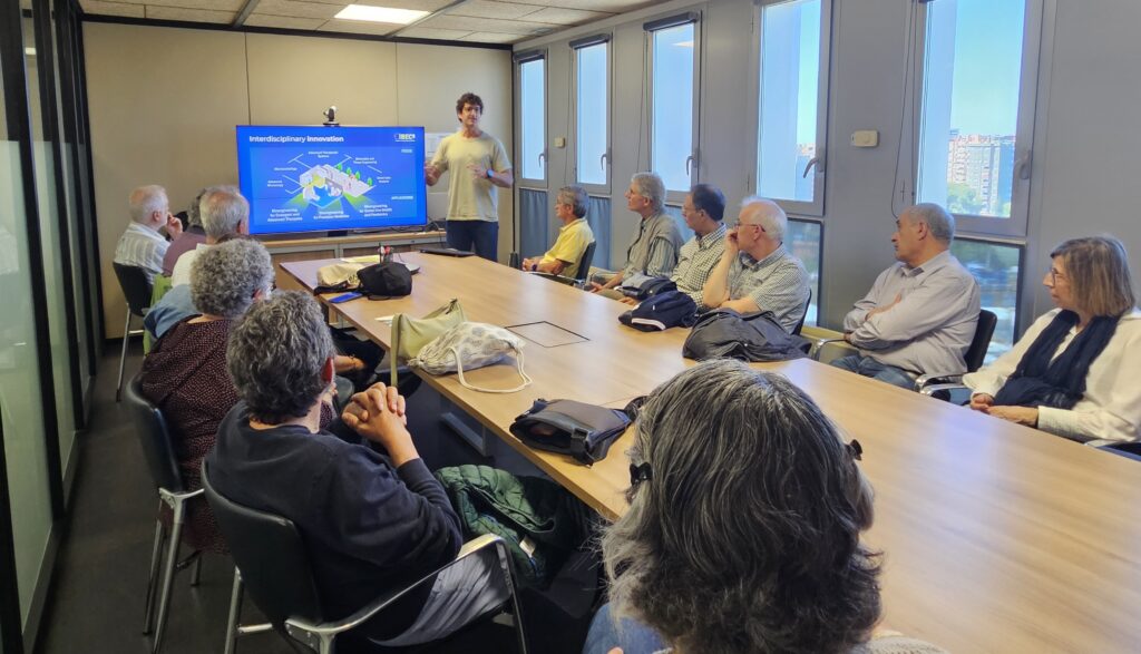
The visit began with a presentation delivered by Gabriel Gomila, leader of the Nanoscale Bioelectrical Characterization group at IBEC and coordinator of the SPM4.0 project. During the session, he introduced the students to the different research areas developed at IBEC before focusing on the work of his own research group and its applications in nanoscale imaging and characterisation.
The presentation provided an overview of several microscopy techniques commonly used in biological and biomedical research, discussing their respective strengths, limitations and suitability for studying different types of samples. This introduction allowed participants to better understand how researchers select the most appropriate imaging approach depending on the scientific question being addressed.
In the second part of the session, Gabriel Gomila presented the fundamentals of Atomic Force Microscopy (AFM), explaining how the technology operates and why it has become a powerful tool for studying living systems. Participants learned how AFM enables researchers to obtain high-resolution information about the structure, mechanical properties and behaviour of biological samples under physiologically relevant conditions. The presentation also included an introduction to the SPM4.0 project and its objectives, highlighting the importance of training a new generation of researchers in advanced scanning probe microscopy technologies.
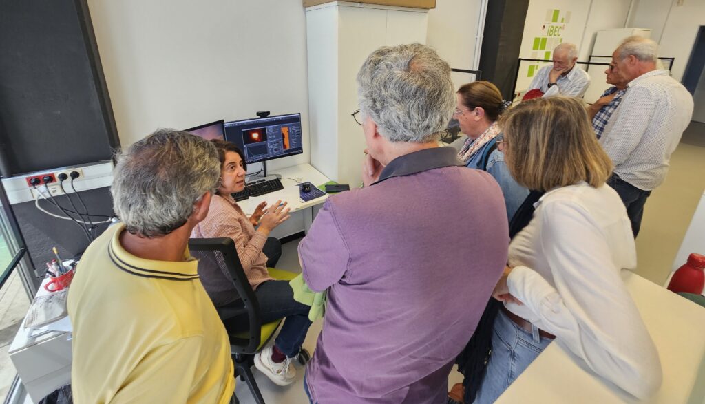
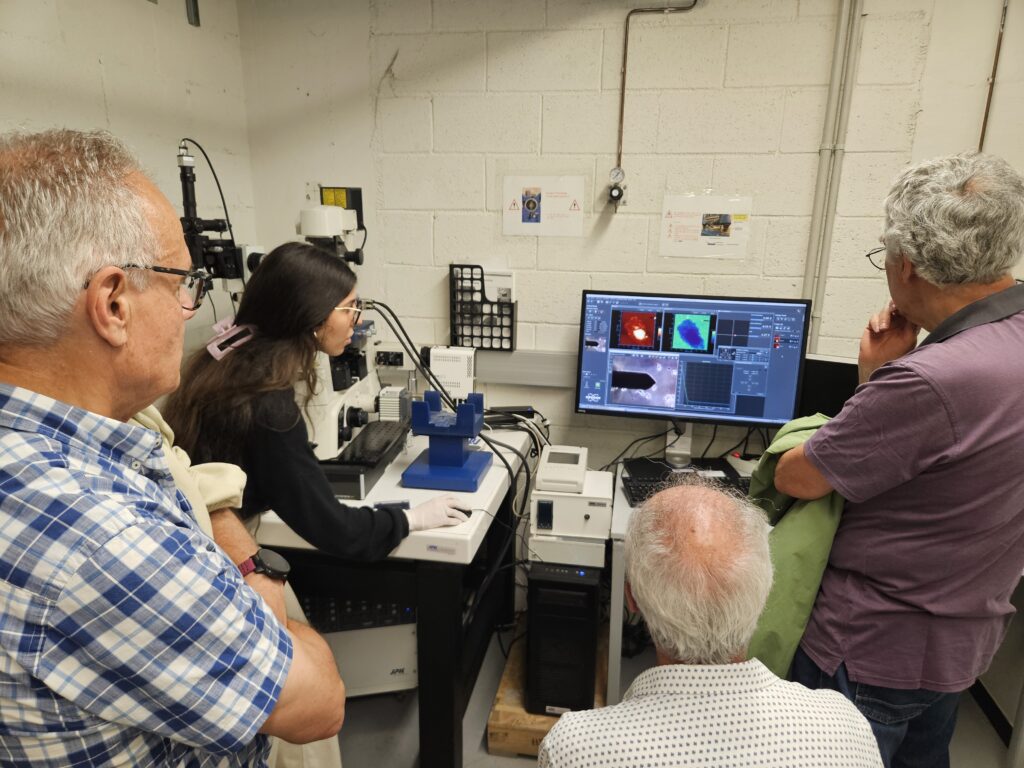
Following the presentation, the students visited the laboratory facilities, where they were guided through three practical stations designed to showcase different aspects of AFM research.
At the first station, with researchers Annalisa Caló and Mauricio Cano Galván, participants explored the software tools used by researchers to process and analyse microscopy images, gaining insight into how raw experimental data are transformed into meaningful scientific information.
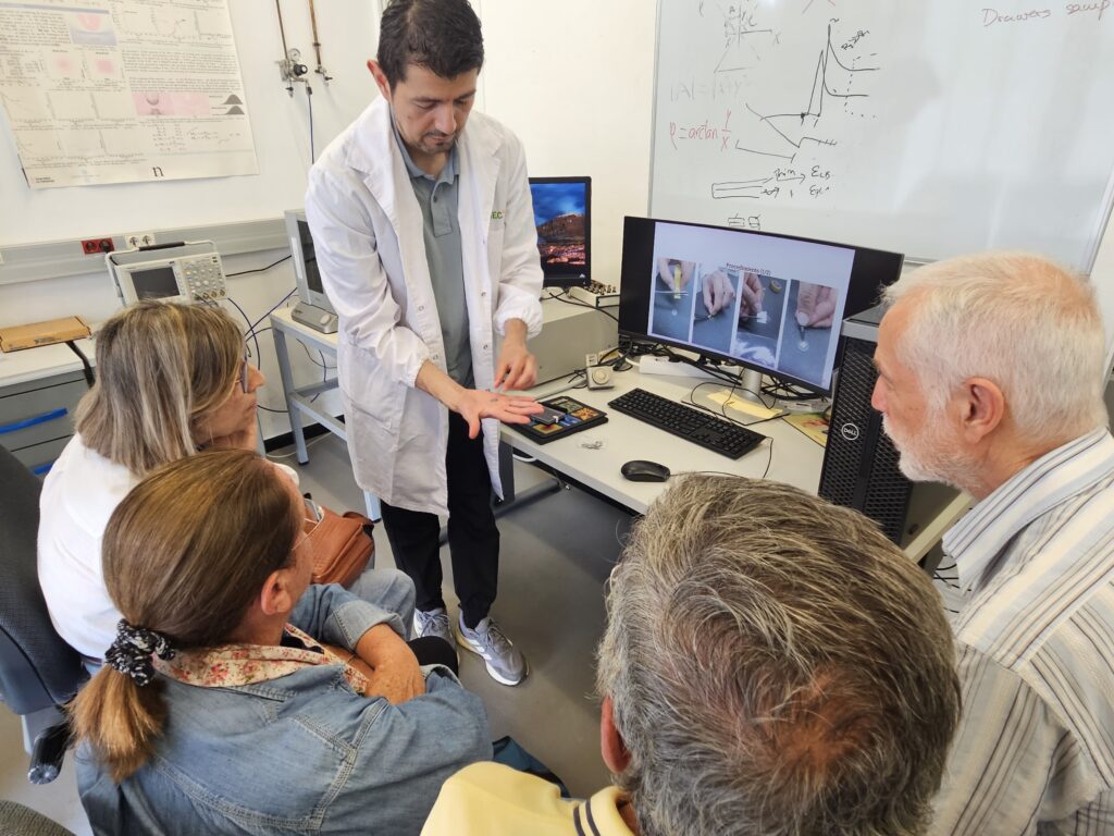
The second station was led by Jhon Pazos, SPM4.0 Doctoral Candidate, who introduced the students to a research-grade AFM instrument. He explained the operating principles of the microscope, sample preparation procedures and the role of key components such as the cantilever and tip. Through real examples, participants learned how different experimental configurations are selected depending on the biological sample under investigation.
The final station focused on a specialised Bio-AFM system adapted for the study of biological specimens. Doctoral researcher Beatriz Cantero demonstrated how this instrument allows scientists to observe and characterise living samples under conditions that closely resemble their natural environment, illustrating the unique capabilities of AFM for biomedical research.
Throughout the visit, students had the opportunity to interact directly with researchers, ask questions and gain a first-hand understanding of the daily activities carried out in a leading bioengineering research institute.
Activities such as this visit to IBEC play an important role in connecting society with scientific research and promoting a greater understanding of emerging technologies. Through the active involvement of researchers such as Gabriel Gomila and Jhon Pazos, the SPM4.0 project continues to showcase the potential of advanced microscopy while inspiring curiosity and interest in science among learners of all ages.